
Topic 1: Cell biology 
This page contains multiple choice questions in the style of Paper 1 of the Biology exams.
They test the breadth of your knowledge of the understandings and skills about cell biology.
To spend more time reviewing the topic before answering these questions, use the revision resources.
Cell biology revision resources
This page lists the understandings and skills expected for Topic 1 and links to the sub-topic pages which contain detailed revision notes, activities and past paper style questions. Great for revision.
Learn from any mistakes. Every question has an examiner's explanation that appears when you check your answers.
Pasteur's experiment with 'swan neck' flasks showed that a sterile nutrient medium exposed to the air would not show any signs of bacterial growth under his conditions.
What prevented the growth of bacteria?
Pasteur's famous experiments with swan neck flasks showed that broth kept in a flask where no dust could settle in the nutrient medium, and thus no living cells could get in, would not go mouldy.
This disproved the theory of spontaeous generation.
The 64 codons in the genetic code give rise to the same amino acids in nearly all organisms.
There is very little variaion. This is evidence for a single common origin of life.
Differences in the frequency of amino acid use reflects the different genes in the two organisms.
Which cells are produced when a diploid human cell divides by mitosis?
Mitosis is division of the nucleus into two genetically identical daughter nuclei in eukaryote cells.
A diploid cell will produce two diploid daughter cells in one division of mitosis.
The four cells shown below have each been surrounded by a solution for 1 hour.
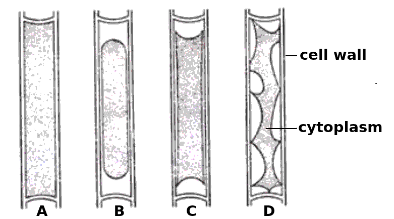
Which cells have been in a hypertonic solution ?
Cell A is swollen turgid, it is in a hypotonic solution or an isotonic solution.
The cells B, C and D show increasing signs of plasmolysis, and so they must be in hypertonic solutions.
The graph below shows the % change in mass of carrot parenchyma slices at different concentrations of sucrose.
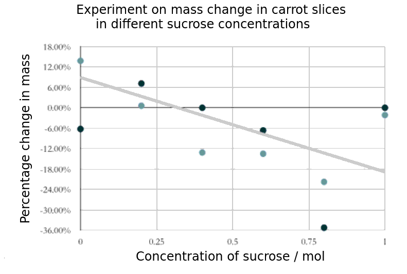
Which of the following is the best estimate of the molarity of the cytoplasm of these cells?
When a sample of cells show no change in mass, then the net movement of water by osmosis must be zero. This shows the concentration of the cytoplasm of the cells. In this graph it would be about 0.3 mol
If you found a eukaryote cell in an electron microscope image, and it contained a lot of rER, Golgi apparatus and many darkly stained vesicles, what do you think the function of the cell is most likely to be?
Cells which make mucus (a protein) will contain lots of rER and vesicles of mucus in secretory vesicles.
Which of the following are methods by which molecules can move across membranes?
- Simple diffusion
- Facilitated diffusion
- Cytokinesis
- Active transport
There are actually four types of membrane transport which are required in DP Biology, Simple diffusion, facilitated diffusion, osmosis and active transport.
The 64 codons in the genetic code give rise to the same amino acids in nearly all organisms.
There is very little variation. This is evidence for a single common origin of life.
Differences in the frequency of amino acid use reflects the different genes in the two organisms.
Which of the following could be used to distinguish a living from a non- living object
Comment: Inanimate objects can move, produce and utilise energy but the process of respiration is exclusive to living systems
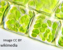
What best describes the organism in the light microscope image?

It is unicellular (one cell) and a eukaryote (has a nucleus) and not autotrophic.
Why is a fungal hypha an exception to the cell theory?

A fungal hypha has many nuclei in a hypha but no cross walls to divide the hypha into cells.
What is the approximate length of the bacterial cell in the image?
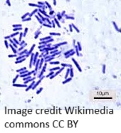
The scale bar is 10 µm and the bacterium is approximately 1 to 1.5 times the length of the scale bar, they are not all the same length.
The image is of a prokaryotic cell. Which feature defines the cell as prokaryotic?
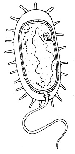
The lack of a membrane bound nucleus (nucleoid) classifies a cell as prokaryotic.
Which organelle is visible in an electron microscope but not in a light microscope?
The ribosome is too small to be seen in the electron microscope, the other organelles were seen in the light microscope before the electron microscope was invented.
The microphotograph is of stratified epithelium. Cells are produced by mitosis in the area marked by the arrow and eventually reach the surface to replace lost cells. Which biological processes does this represent?
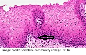
The cells produced by mitosis differentiate into mature cells and replace the cells lost at the surface.
Which means of transport across a plasma membrane requires the molecule shown in the picture?

What tonicity should a saline drip for rehydration have in comparison to human blood?
The saline drip must be isotonic to human blood to not cause water gain or loss from tissues.
Which component of the plasma membrane varies in function between differentiated cells?
The protein component of the membrane varies in structure and function.
Which of the following contributed to the acceptance of the fluid mosaic model of membrane structure of Singer and Nicholson in place of the original Davison-Danielli model?
I Hydrophobic membrane proteins
II Irregular sizes of membrane proteins
III Increased magnification of light microscopes.
IV Fluorescent antibody tagging.
The irregular sizes and insolubility of hydrophobic membrane proteins indicated that they could not be a surface layer as proposed by Davison-Danielli. This was confirmed by fluoresecent antibodies showing that proteins were both within and on the membrane.
The image below was taken in 1825 and shows part of the cell cycle.
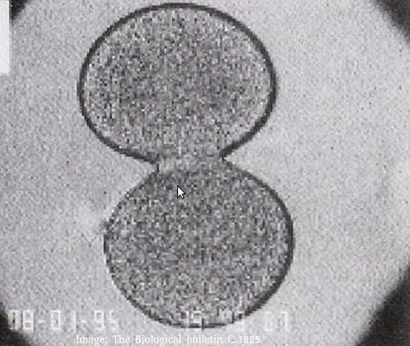
What is shown in the image?
Cytokinesis occurs after mitosis in plant and animal cells.
Animal cells form a cleavage furrow (looks like a wasps waist) as they don't have cell walls.
The two daughter cells are the same size, so cytokinesis is equal.
Refresh this page to try a new set of 20 multiple choice questions. The questions will be different next time you visit. Great revision.
 Twitter
  Facebook
  LinkedIn