
Topic 1: Cell biology 
This page contains multiple choice questions in the style of Paper 1 of the Biology exams.
They test the breadth of your knowledge of the understandings and skills about cell biology.
To spend more time reviewing the topic before answering these questions, use the revision resources.
Cell biology revision resources
This page lists the understandings and skills expected for Topic 1 and links to the sub-topic pages which contain detailed revision notes, activities and past paper style questions. Great for revision.
Learn from any mistakes. Every question has an examiner's explanation that appears when you check your answers.
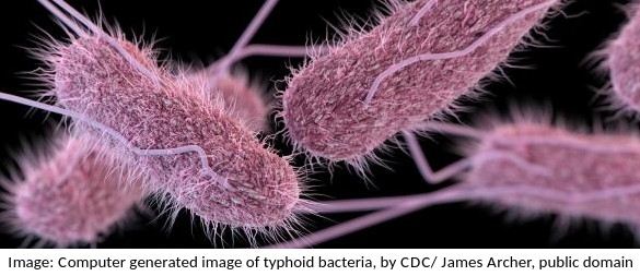
Pasteur's experiment with 'swan neck' flasks showed that a sterile nutrient medium exposed to the air would not show any signs of bacterial growth under his conditions.
What prevented the growth of bacteria?
Pasteur's famous experiments with swan neck flasks showed that broth kept in a flask where no dust could settle in the nutrient medium, and thus no living cells could get in, would not go mouldy.
This disproved the theory of spontaeous generation.
The image below was taken in 1825 and shows part of the cell cycle.
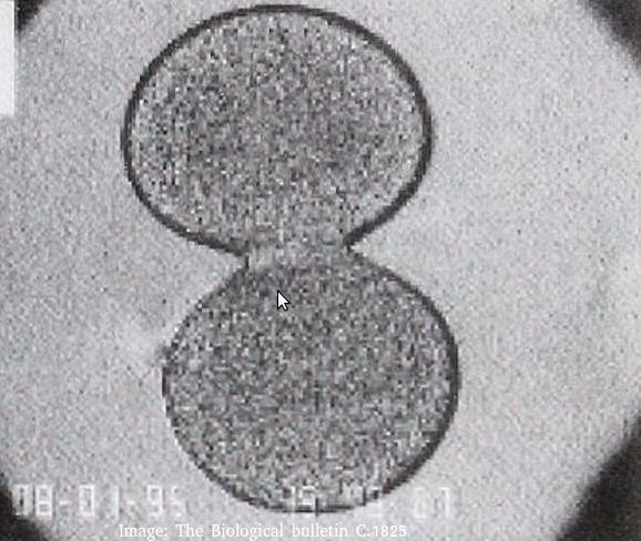
What type of cells is this and at which stage of the cell cycle?
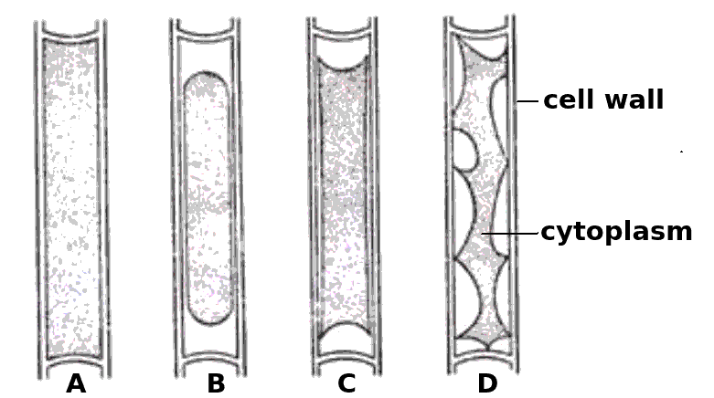
Cytokinesis occurs after mitosis in plant and animal cells. The chromosomes are uncoiled.
Plant cells build a new cell wall which divides the cytoplasm.
Animal cells form a cleavage furrow (likes a wasps waist) as they don't have cell walls.
Which one of the statements below best describes the mitotic index?
The mitotic shows the speed of cell division, which can be used as a tool to identify cancer.
It is calculated by dividing the number of cells doing mitosis by the total number of cells.
The four cells shown below have each been surrounded by a solution for 1 hour.
Which cells have been in a hypertonic solution ?
Cell A is swollen turgid, it is in a hypotonic solution or an isotonic solution.
The cells B, C and D show increasing signs of plasmolysis, and so they must be in hypertonic solutions.
Skill: Estimation of osmolarity in tissues by bathing samples in hypotonic and hypertonic solutions. (Practical 2)
The electron microscope image below shows a ciliated epithelial cell from the trachea.
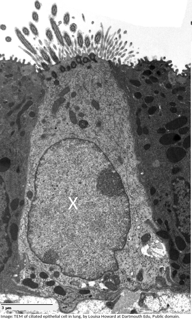
What is the name of the organelle labelled X?
What are the structures labelled X and Y likely to be in this electron microscope image?
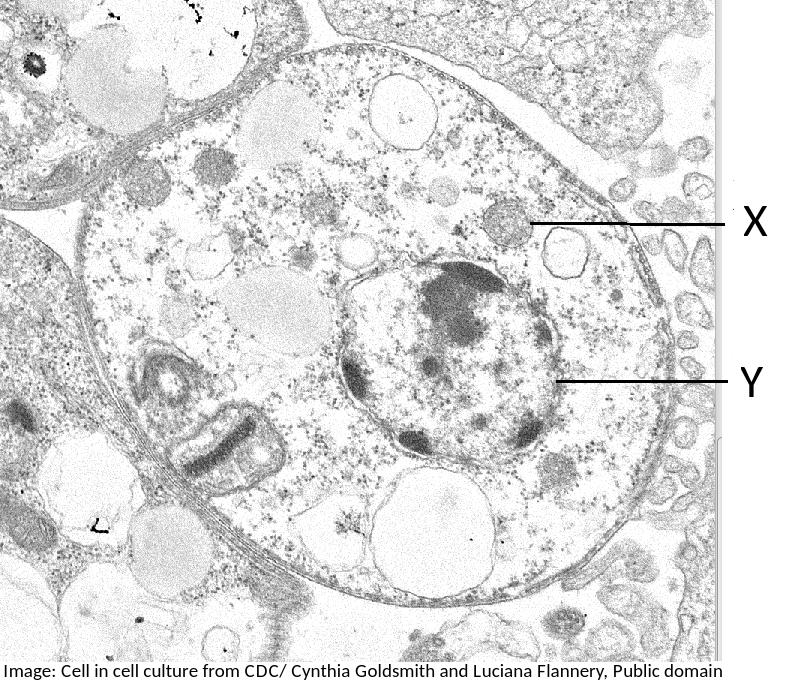
Students are expected to be able to identify organelles from microscope images of cells. The nucleus is distinctive because it is about 10µm in size, and it has black dots in it, chromatin, and sometimes one or more dark patches within the nuclear membrane. It also has a double membrane, not often easily visible.
This electron microscope image shows a group of prokaryotes.
What structures are most likely to be found inside these cells?
Skill: you should know how to draw prokaryotic cells (with a cell wall, plasma membrane, cytoplasm, pili, flagella, 70s ribosomes and nucleoid.) and eukaryotic cells (free 80s ribosomes, rough endoplasmic reticulum (rER), lysosome, Golgi apparatus, mitochondrion and nucleus)
Cell theory covers most, but not all cases.
Which one of these statements is an exception?
Exceptions to cell theory are : multinucleated striated muscle the giant single celled Acetabularia algae?
Also, organisms consisting of only one cell carry out all functions of life in that cell. e.g. Paramecium, Chlorella.
What is the importance of surface area to volume ratio to cells?
Surface area to volume ratio is important in the limitation of cell size. The lager the volume, the greater the need for materials which have to be exchanged over the surface of the cell.
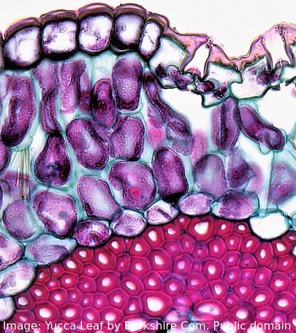 The image shows a range of different cell types in the leaf of a Yucca plant.
How do stem cells form this range of cells?
What is the process called?
Specialised tissues can develop by cell differentiation in multicellular organisms.
Differentiation involves the expression of some genes and not others in a cell.
The blood cells below were imaged using an electron microscope.
The magnification is x3000 and the ruler measures the central cell as being 2 cm in diameter.
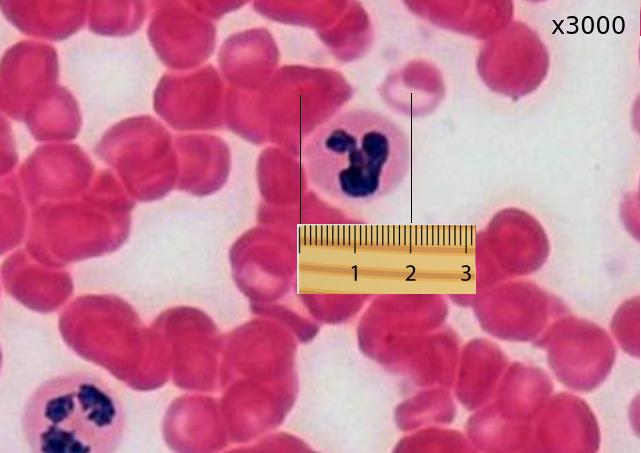
Estimate the actual size of this white blood cell.
Calculate specimen size using magnification?
First change the size measurement into µm units = 20000µm
Then divide by the magnification. 20000 / 3000 = 20 / 3 = 6.6 µm
Human red blood cells are circular and 0.6 μm in diameter. A photograph of a red blood cell is shown as an illustration in a book with a diameter of 1.2mm. What is the magnification of the diagram?
Comment: Convert 1.2 mm into μm by multiplying x 1000 = 1200 μm (so that both units are the same). Then you can see that 0.6 x 2000 = 1200. Or use the formula Magnification = Image size/true size. If the photograph is larger than the cell, the magnification could not be 0.5x which would make it smaller. Eliminate obviously incorrect answers.
The image below is of Dracaena leaf upper epidermis cells.
Which of the following is the best estimate of the length (from top to bottom) of an epidermal cell?
Comment: The cells are approximately the same size as the scale bar.
This would make 70µm the closest estimate.
Which of the following are methods by which molecules can move across membranes?
I. Simple diffusion
II. Facilitated diffusion
III. Cytokinesis
IV. Active transport
There are actually four types of membrane transport which are required in DP Biology, Simple diffusion, facilitated diffusion, osmosis and active transport.
Which organelle in a eukarytotic animal cell synthesises proteins for exocytosis?

The RER synthesises proteins for exocytosis.
The diagram is of a plasma membrane. Which label corresponds to an extrinsic glycoprotein?

Extrinsic proteins are on the outside of the membrane, glycoproteins have carbohydrate prosthetic (side) groups (shown by the hexagonal shape).
All eukaryote cells have differing combinations of the same types of organelles. How can this be explained?
Similarity in structure and function are taken as evidence of a common ancestor. All of the other statements are partially true but do not provide an explanation.
Which of the following is the best description of an organelle?
The "wrong" answers are correct statements but are distractors, not the best description.
Why is the cell component in the image regarded as an organelle?

The organelle is a cell component with a membane, the mitochondrion, it is adapted to aerobic respiration.
There are twenty complete cells in this microphotograph (with complete nuclear material). Estimate the number of complete cells in prophase of mitosis.

There are 3 complete cells in prophase (chromosomes visible in a nucleus..
Refresh this page to try a new set of 20 multiple choice questions. The questions will be different next time you visit. Great revision.
 Twitter
  Facebook
  LinkedIn