
Topic 1: Cell biology 
This page contains multiple choice questions in the style of Paper 1 of the Biology exams.
They test the breadth of your knowledge of the understandings and skills about cell biology.
To spend more time reviewing the topic before answering these questions, use the revision resources.
Cell biology revision resources
This page lists the understandings and skills expected for Topic 1 and links to the sub-topic pages which contain detailed revision notes, activities and past paper style questions. Great for revision.
Learn from any mistakes. Every question has an examiner's explanation that appears when you check your answers.
Huntington's Disease (HD) is a brain disorder that affects a person's ability to talk, and move. HD is caused by a faulty protein. The job of the protein is to direct vesicles containing important molecules to the outside of the cell. The chemicals are released when the vesicle reaches the membrane.
What is the name given to the release of chemicals by a cell in this way?
This is a type of secretion, the vesicle fuses with the plasma membrane and its contents are released. This is called "exocytosis".
Cells today come from pre-existing cells. The origin of the fist cell must be different.
Where do biologists think the first cell came from?
The first cell must have come from non-living material. This material must have contained molecules which today we consider as organic, carbon containing molecules.
The image below shows a eukaryotic cell.
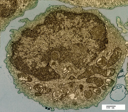
Which structure, visible in the image, could be used as evidence supporting endosymbiosis?
The mitochondria provide evidence supporting endosymbiosis because they have:
- a double membrane
- 70S ribosomes
- DNA
The image below was taken in 1825 and shows part of the cell cycle.
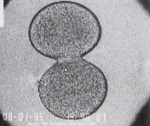
What type of cells is this and at which stage of the cell cycle?
Cytokinesis occurs after mitosis in plant and animal cells. The chromosomes are uncoiled.
Plant cells build a new cell wall which divides the cytoplasm.
Animal cells form a cleavage furrow (likes a wasps waist) as they don't have cell walls.
During interphase of the cell cycle what happens to the DNA in the nucleus?
Under the microscope there is little change during interphase.
However interphase is a very active phase of the cell cycle with many processes occurring in the nucleus and cytoplasm. (It is subdivided into G1, S, G2 phases of the cell cycle)
DNA replication occurs during the S-phase.
The four cells shown below have each been surrounded by a solution for 1 hour.
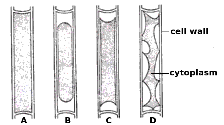
Which cells have been in a hypertonic solution ?
Cell A is swollen turgid, it is in a hypotonic solution or an isotonic solution.
The cells B, C and D show increasing signs of plasmolysis, and so they must be in hypertonic solutions.
Skill: Estimation of osmolarity in tissues by bathing samples in hypotonic and hypertonic solutions. (Practical 2)
Stargardts disease is vision loss caused by the death of both cone cells and rod cells in the part of the retina around the fovea. One potential treatment for Stargardts disease is the use of human embryonic stem cells.
What are the properties of these stem cells, which other cells don't have, that makes them so useful for this treatment?
Stem cells can divide and this help researchers to grow them in the lab.
They can differentiate along different pathways in embryonic development which makes stem cells useful for therapeutic uses (e.g. Stargart's disease) because they can be grown into many different tissues.
The electron microscope image below shows a scale bar marked with 100µm.
The large 'goblet cell' in the centre is producing mucous which will protect the surface of the epithelium.
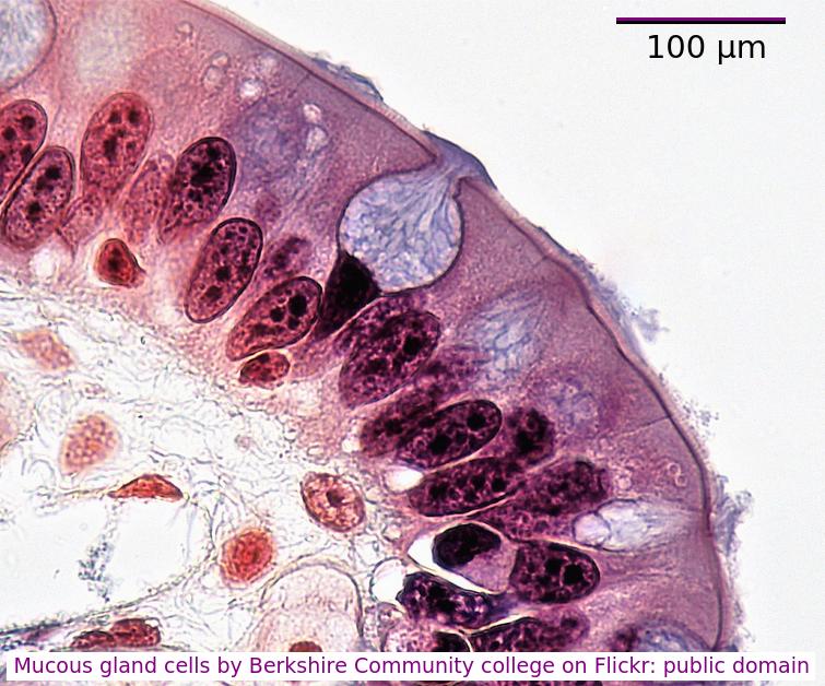
What is the diameter of the goblet cell?
Accurately, measure the scale bar length in mm, measure the diameter of the cell, in mm
divide cell diameter by scalebar and multiply by 100µm.
You can often estimate the size using the scale bar and your thumb or a pen.
Which label in the image below shows the process of endocytosis?
![]()
Endocytosis is the process where a substance is surrounded by the plasma membrane which forms a vesicle inside the cell that then moves into the cytoplasm, separating from the plasma membrane.
Which of the following are methods by which molecules can move across membranes?
I. Simple diffusion
II. Facilitated diffusion
III. Cytokinesis
IV. Active transport
There are actually four types of membrane transport which are required in DP Biology, Simple diffusion, facilitated diffusion, osmosis and active transport.
The diagram shows a typical eukaryotic plant cell. Which organelles are involved in supporting the cell and plant? I Cell wall II Cytoplasm III Nucleus IV Vacuole.

The cell and plant are supported by the turgor pressure of water in the vacuole acting on the rigid cell wall.
Which organelles are found in large numbers in secretory cells in animals? I Vesicles II Golgi Body III Mitochondria IV Rough endoplasmic reticulum.

Secretory cells synthesise proteins for exocytosis so have large numbers of mitochondria to supply energy, RER to synthesise the proteins for packaging into vesicles by the Golgi Body.
The diagram is of a plasma membrane. Which label corresponds to a protein channel?
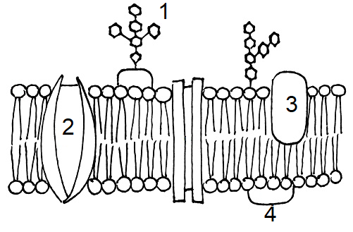
Protein channels cross the membrane to allow hydrophilic substances to pass through the membrane.
The diagram is of a plasma membrane. Which label corresponds to the hydrophilic area of an amphipathic molecule?

Protein (5) has both hydrophilic and hydrophobic areas to act as an integral protein. The central channel is hydrophilic.
The image is a ribbon model of a channel protein. Where would this be found in a plasma membrane?

Channel proteins penetrate the membrane and have a central hydrophilic area (yellow in the diagram which is shown from above).
Which process is involved in white blood cells engulfing bacteria (arrowed in the photomicrograph)?

White blood cells engulf bacteria by endocytosis, the intake of solid particles by a cell membrane.
Which organelles in a plant cell are believed to have originated as free-living prokaryotic cells?

Both the mitochondria and the chloroplast in plant cells are thought to have been free-living prokaryotes which evolved in a symbiotic relationship with a eukaryotic cell.
Identify the stage of mitosis of cells 1 and 2
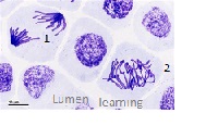
In cell 2, the chromatids are aligned on the equator (seen from above)- Metaphase. In cell 1, the chromatids are moving towards the poles - Anaphase.
Which component of the plasma membrane varies in function between differentiated cells?
The protein component of the membrane varies in structure and function.
Which of the following contributed to the acceptance of the fluid mosaic model of membrane structure of Singer and Nicholson in place of the original Davison-Danielli model?
I Hydrophobic membrane proteins
II Irregular sizes of membrane proteins
III Increased magnification of light microscopes.
IV Fluorescent antibody tagging.
The irregular sizes and insolubility of hydrophobic membrane proteins indicated that they could not be a surface layer as proposed by Davison-Danielli. This was confirmed by fluoresecent antibodies showing that proteins were both within and on the membrane.
Refresh this page to try a new set of 20 multiple choice questions. The questions will be different next time you visit. Great revision.
 Twitter
  Facebook
  LinkedIn