
Topic 1: Cell biology 
This page contains multiple choice questions in the style of Paper 1 of the Biology exams.
They test the breadth of your knowledge of the understandings and skills about cell biology.
To spend more time reviewing the topic before answering these questions, use the revision resources.
Cell biology revision resources
This page lists the understandings and skills expected for Topic 1 and links to the sub-topic pages which contain detailed revision notes, activities and past paper style questions. Great for revision.
Learn from any mistakes. Every question has an examiner's explanation that appears when you check your answers.
What is the best definition of endosymbiosis?
Endosymbiosis is where a cell engulfs another cell and it continues to live inside the cell.
The engulfed cell provides something for the host cell, and gets something in return. Both cells benefit.
Which cells are produced when a diploid cell divides by mitosis?
Mitosis is division of the nucleus into two genetically identical daughter nuclei in eukaryote cells.
A diploid cell will produce two diploid daughter cells in one division of mitosis.
The DNA of eukaryote cells is organised into chromosomes
What happens to the DNA at prophase in the beginning of mitosis?
Chromosomes condense by supercoiling during mitosis. This makes the chromosomes visible.
The DNA replicates during interphase, not prophase.
Which one of the statements below best describes the mitotic index?
The mitotic shows the speed of cell division, which can be used as a tool to identify cancer.
It is calculated by dividing the number of cells doing mitosis by the total number of cells.
What effect does reducing the amount of cholesterol in a cell membrane have on its properties?
Cholesterol is a component of animal cell membranes. Application: Cholesterol in mammalian membranes reduces membrane fluidity and permeability to some solutes.
The image below shows three structures as seen in an electron microscope.
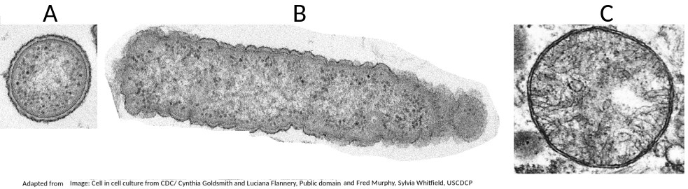
Which of the structures are prokaryote cells?
Students are expected to be able to recognise and draw the simple structure of Prokaryote cells.
There is no compartmentation in prokaryote cells, and as membranes can be seen in structure B (a mitochondrion) it is not a prokaryote.
The electron microscope image below shows an organelle found in both animal and plant cells.
What is the name of the organelle?
Know how to idenfity the organelles in eukaryotes and draw their compartmentalised structure.
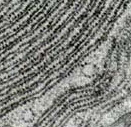
The rER has parallel membranes covered in dots, which are ribosomes, used for making proteins, for secretion from the cell.
What are the structures labelled X and Y likely to be in this electron microscope image?
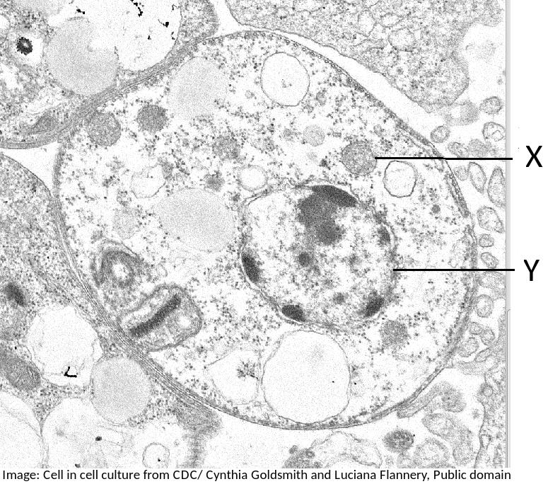
Students are expected to be able to identify organelles from microscope images of cells. The nucleus is distinctive because it is about 10µm in size, and it has black dots in it, chromatin, and sometimes one or more dark patches within the nuclear membrane. It also has a double membrane, not often easily visible.
The electron microscope image below shows a cell.
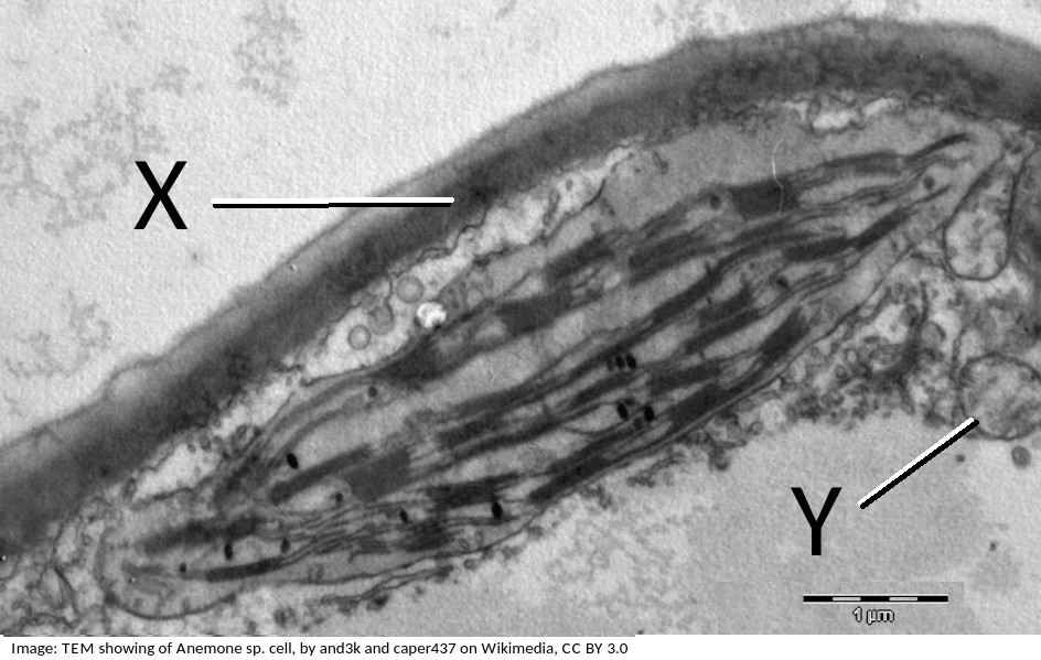
What are the organelles shown by the labels X & Y?
If you look closely at X, it points to the cell wall, outside the plasma membrane, it is close to the plasma membrane, but not touching the chloroplast.
The pale area below Y is the vacuole.
Organelle Y is a mitochondrion, you can tell this by its size, and the presence of membranes inside.
When nerve cells form brain tissue they can; store memories, create thoughts and coordinate movement.
If you only ever studied individual nerve cells you would never see these abilities which the brain has.
What is this type of property called?
Multicellular organisms have properties that emerge from the interaction of their cellular components. (Emergent properties)
Stargardts disease is vision loss caused by the death of both cone cells and rod cells in the part of the retina around the fovea. One potential treatment for Stargardts disease is the use of human embryonic stem cells.
What are the properties of these stem cells, which other cells don't have, that makes them so useful for this treatment?
Stem cells can divide and this help researchers to grow them in the lab.
They can differentiate along different pathways in embryonic development which makes stem cells useful for therapeutic uses (e.g. Stargart's disease) because they can be grown into many different tissues.
Which label in the image below shows the process of endocytosis?
![]()
Endocytosis is the process where a substance is surrounded by the plasma membrane which forms a vesicle inside the cell that then moves into the cytoplasm, separating from the plasma membrane.
Which is the correct order of SI units, beginning with the largest?
Comment: SI units always have a differential of 1000. The unit without the prefix is the standard SI unit (metre, m). B and D are clearly wrong, eliminate those answers first.
The image below is of Dracaena leaf upper epidermis cells.
Which of the following is the best estimate of the length (from top to bottom) of an epidermal cell?
Comment: The cells are approximately the same size as the scale bar.
This would make 70µm the closest estimate.
Why is a fungal hypha an exception to the cell theory?

A fungal hypha has many nuclei in a hypha but no cross walls to divide the hypha into cells.
Which process is involved in white blood cells engulfing bacteria (arrowed in the photomicrograph)?

White blood cells engulf bacteria by endocytosis, the intake of solid particles by a cell membrane.
What is the sequence of events that occur in a cell that is secreting a protein hormone?
1 Exocytosis
2 Vesicle formed by Golgi Body
3 Fusion of vesicle to plasma membrane
4 RER manufactures protein.
Ribosomes on the RER manufacture protein. This is packaged in vesicles by the Golgi Body and moves to the surface of the cell where the vesicle and plasma membrane fuse and exocytosis of the protein occurs.
Which of the following contributed to the acceptance of the fluid mosaic model of membrane structure of Singer and Nicholson in place of the original Davison-Danielli model?
I Hydrophobic membrane proteins
II Irregular sizes of membrane proteins
III Increased magnification of light microscopes.
IV Fluorescent antibody tagging.
The irregular sizes and insolubility of hydrophobic membrane proteins indicated that they could not be a surface layer as proposed by Davison-Danielli. This was confirmed by fluoresecent antibodies showing that proteins were both within and on the membrane.
Which of the following is the best description of an organelle?
The "wrong" answers are correct statements but are distractors, not the best description.
A tissue is placed in an isotonic solution. Which of the following is the best description of water movement between the tissue and the solution?
There is no net water movement, gain and loss from the tissue is equal in both directions.
Refresh this page to try a new set of 20 multiple choice questions. The questions will be different next time you visit. Great revision.
 Twitter
  Facebook
  LinkedIn