
Topic 1: Cell biology 
This page contains multiple choice questions in the style of Paper 1 of the Biology exams.
They test the breadth of your knowledge of the understandings and skills about cell biology.
To spend more time reviewing the topic before answering these questions, use the revision resources.
Cell biology revision resources
This page lists the understandings and skills expected for Topic 1 and links to the sub-topic pages which contain detailed revision notes, activities and past paper style questions. Great for revision.
Learn from any mistakes. Every question has an examiner's explanation that appears when you check your answers.
Huntington's Disease (HD) is a brain disorder that affects a person's ability to talk, and move. HD is caused by a faulty protein. The job of the protein is to direct vesicles containing important molecules to the outside of the cell. The chemicals are released when the vesicle reaches the membrane.
What is the name given to the release of chemicals by a cell in this way?
This is a type of secretion, the vesicle fuses with the plasma membrane and its contents are released. This is called "exocytosis".
The image below shows a eukaryotic cell.
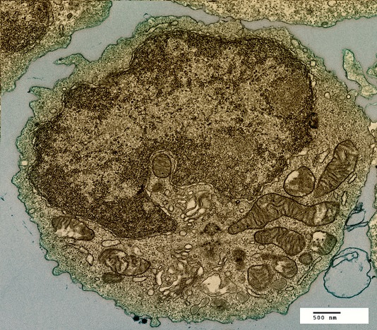
Which structure, visible in the image, could be used as evidence supporting endosymbiosis?
The mitochondria provide evidence supporting endosymbiosis because they have:
- a double membrane
- 70S ribosomes
- DNA
The image below shows two cells undergoing mitosis.
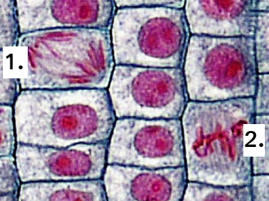
Which of the following correctly identifies the stage of mitosis for each of the cells?
Identification of the phase of mitosis is an important skill.
The position of the chromosomes gives the best clue.
In Prophase, they are spread around the cell, and double stranded.
In Metaphase, the chromosomes are lined up on the spindle equator.
In Anaphase, the chromosomes are moving to opposite poles of the cell.
In Telophase, the two groups of chromosomes are separate, sometimes with a nuclear membrane.
The illustration shown below is of a protein (green) attached to a membrane.
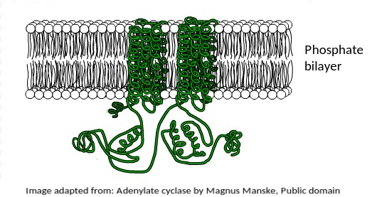
What is the most likely function of this membrane protein?
This protein is found in the human liver, where the hormone adrenaline indirectly stimulates it to mobilise stored energy inside liver cells in the "fight or flight" response.
The fact that it is a transmembrane protein is essential for this function.
It is interesting to note that this protein is also secreted by Anthrax bacteria as a toxin.
It upsets the metabolism of host cells when it enters them.
The electron microscope image below shows an organelle found in eukaryote cells.
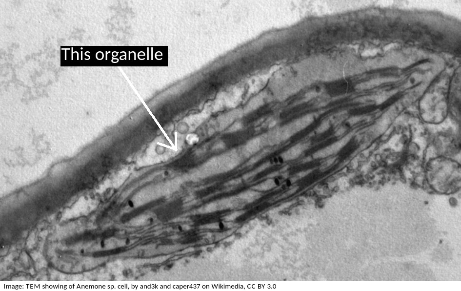
What is the name of the organelle?
Chloroplasts are distinctive because they have stacks of membranes inside, called grana, which hold the chlorophyll that absorbs light.
How does compartmentalisation by their internal membranes benefit eukaryotic cells?
Eukaryote cells (approx. 100µm in diameter) are much larger than prokaryote cells (approx 1µm) and so the concentration of reactants in the cytoplasm would be more dilute if all the metabolism happened in the cytoplasm.
Specialist organelles, like mitochondria keep the enzymes for aerobic respiration in one place, which increases their concentration, and increases the rate of reactions.
Cell theory covers most, but not all cases.
Which one of these statements is an exception?
Exceptions to cell theory are : multinucleated striated muscle the giant single celled Acetabularia algae?
Also, organisms consisting of only one cell carry out all functions of life in that cell. e.g. Paramecium, Chlorella.
Which of the following are methods by which molecules can move across membranes?
- Simple diffusion
- Facilitated diffusion
- Cytokinesis
- Active transport
There are actually four types of membrane transport which are required in DP Biology, Simple diffusion, facilitated diffusion, osmosis and active transport.
Which of the structures listed below are involved in membrane transport?
Many transmembrane proteins are involved in transport of molecules across membranes. These can either provide a sort of molecular pore through which ions or molecules can pass (facilitated diffusion), or they can use ATP to actively move molecules, even against the concentration gradient (active transport).These are just two examples, transport can also occur by simple diffusion through the phospholipid bilayer, or by endocytosis.
Cells are often stored in isotonic conditions because they can be damaged in other concentrations, hypertonic, or hypotonic. Which of the descriptions of hypertonic is the most accurate?
Hypertonic solutions have a higher concentration of solutes, and lower water potentials than cells.
Which two processes are involved in cell differentiation?
Comment: During differentiation genes may be activated or repressed causing the formation of different proteins by the ribosomes but other organelles such as mitochondria continue to function in the same way
Which of the following could be used to distinguish a living from a non- living object
Comment: Inanimate objects can move, produce and utilise energy but the process of respiration is exclusive to living systems
Human red blood cells are circular and 0.6 μm in diameter. A photograph of a red blood cell is shown as an illustration in a book with a diameter of 1.2mm. What is the magnification of the diagram?
Comment: Convert 1.2 mm into μm by multiplying x 1000 = 1200 μm (so that both units are the same). Then you can see that 0.6 x 2000 = 1200. Or use the formula Magnification = Image size/true size. If the photograph is larger than the cell, the magnification could not be 0.5x which would make it smaller. Eliminate obviously incorrect answers.
Which is the correct order of SI units, beginning with the largest?
Comment: SI units always have a differential of 1000. The unit without the prefix is the standard SI unit (metre, m). B and D are clearly wrong, eliminate those answers first.
The diameter of this field of view under a microscope at X400 magnification is 250 μm.
The image below shows Dracaena leaf upper epidermis cells.
Which is the best estimate of the width (from left to right) of an epidermal cell?
Comment: There are approximately 8 cells across a diameter so 250/8 = 33 μm.
Beware of the units, mm are 1000 times bigger than µm.
The image is a ribbon model of a channel protein. Where would this be found in a plasma membrane?

Channel proteins penetrate the membrane and have a central hydrophilic area (yellow in the diagram which is shown from above).
Which process is involved in white blood cells engulfing bacteria (arrowed in the photomicrograph)?

White blood cells engulf bacteria by endocytosis, the intake of solid particles by a cell membrane.
What is the sequence of events that occur in a cell that is secreting a protein hormone?
1 Exocytosis
2 Vesicle formed by Golgi Body
3 Fusion of vesicle to plasma membrane
4 RER manufactures protein.
Ribosomes on the RER manufacture protein. This is packaged in vesicles by the Golgi Body and moves to the surface of the cell where the vesicle and plasma membrane fuse and exocytosis of the protein occurs.
A mitotic index taken from this microphotograph only would not be regarded as valid. How can a valid count be made?

Which of the following are believed to be endosymbiotic structures involved in cell locomotion in both prokaryotes and eukaryotes?
Flagellae are locomotory structures found in some Monera (bacteria), and some eukaryotic cells such as male gametes and Protoctista. Mitochondria are not found in prokaryotes. Pseudopodia are involved in locomotion but only in cells without an external wall. Fimbriae in bacteria allow for binding to a host or substrate, the same name is given to projections in the oviduct that aid movement of the ovum towards the uterus.
Refresh this page to try a new set of 20 multiple choice questions. The questions will be different next time you visit. Great revision.
 Twitter
  Facebook
  LinkedIn