
Topic 1: Cell biology 
This page contains multiple choice questions in the style of Paper 1 of the Biology exams.
They test the breadth of your knowledge of the understandings and skills about cell biology.
To spend more time reviewing the topic before answering these questions, use the revision resources.
Cell biology revision resources
This page lists the understandings and skills expected for Topic 1 and links to the sub-topic pages which contain detailed revision notes, activities and past paper style questions. Great for revision.
Learn from any mistakes. Every question has an examiner's explanation that appears when you check your answers.
Huntington's Disease (HD) is a brain disorder that affects a person's ability to talk, and move. HD is caused by a faulty protein. The job of the protein is to direct vesicles containing important molecules to the outside of the cell. The chemicals are released when the vesicle reaches the membrane.
What is the name given to the release of chemicals by a cell in this way?
This is a type of secretion, the vesicle fuses with the plasma membrane and its contents are released. This is called "exocytosis".
The 64 codons of mRNA code for the same amino acids in almost all species. A rare exception is found in Paramecium where one of the "stop codons" actually codes for the amino acid glutamine.
What does this suggest about the origin of cells?
The 64 codons in the genetic code give rise to the same amino acids in nearly all organisms, There is very little variaion. If the genetic code had evolved several times in the history off life, there would be many differences.
Which property of phospholipid molecules describes the fact that they have both hydrophobic and hydrophilic parts?
Phospholipids form bilayers in water due to the amphipathic properties of phospholipid molecules. The hydrophobic tails attract each other and the hydrophilic phosphates are attracted to the water.
Which of the following is true of peripheral proteins in cell membranes?
Membrane proteins are diverse in terms of structure, position in the membrane and function.
Peripheral proteins are attached to the membrane but found on its surface.
The illustration shown below is of a protein (green) attached to a membrane.
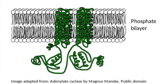
What is the most likely function of this membrane protein?
This protein is found in the human liver, where the hormone adrenaline indirectly stimulates it to mobilise stored energy inside liver cells in the "fight or flight" response.
The fact that it is a transmembrane protein is essential for this function.
It is interesting to note that this protein is also secreted by Anthrax bacteria as a toxin.
It upsets the metabolism of host cells when it enters them.
The electron microscope image below shows an organelle found in both animal and plant cells.
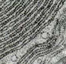
What is the name of the organelle?
Know how to idenfity the organelles in eukaryotes and draw their compartmentalised structure.
The rER has parallel membranes covered in dots, which are ribosomes, used for making proteins, for secretion from the cell.
The electron microscope image below shows an organelle found in eukaryote cells.
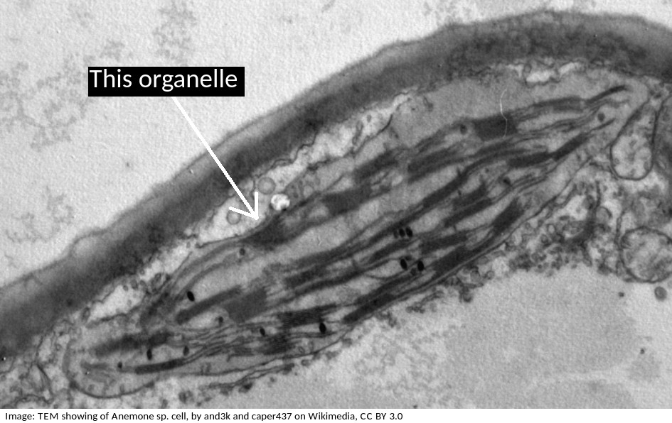
What is the name of the organelle?
Chloroplasts are distinctive because they have stacks of membranes inside, called grana, which hold the chlorophyll that absorbs light.
This electron microscope image shows a group of prokaryotes.
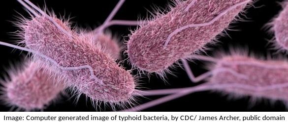
What structures are most likely to be found inside these cells?
Skill: you should know how to draw prokaryotic cells (with a cell wall, plasma membrane, cytoplasm, pili, flagella, 70s ribosomes and nucleoid.) and eukaryotic cells (free 80s ribosomes, rough endoplasmic reticulum (rER), lysosome, Golgi apparatus, mitochondrion and nucleus)
Cell theory covers most, but not all cases.
Which one of these statements is an exception?
Exceptions to cell theory are : multinucleated striated muscle the giant single celled Acetabularia algae?
Also, organisms consisting of only one cell carry out all functions of life in that cell. e.g. Paramecium, Chlorella.
The image below shows erythrocytes and leucocytes.l.
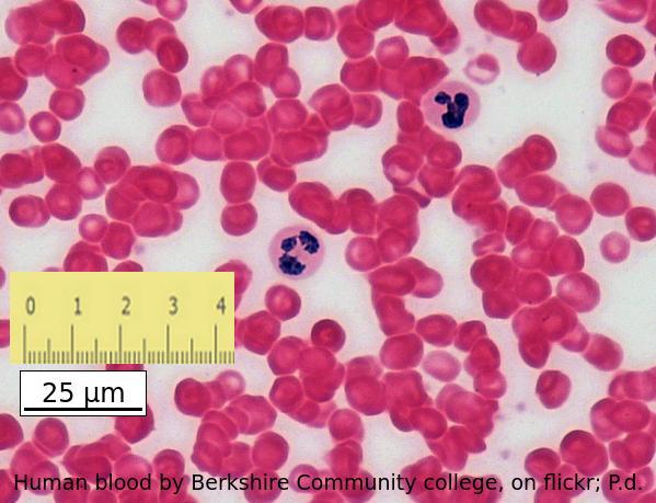
Using the scale bar and the ruler placed on the image, estimate the magnification of the image.
Which answer is the best estimate
Calculate the magnification of an electron microscope image from a scale bar?
Convert the ruler measurement to the same units written on the scale bar, in this case 25mm is 25000µm
then divide the ruler measurement 25000 by the number on the scalebar, 25.
Stargarts disease, which causes loss of cells in the retina can be treated using a special type of human cell.
Which of the following is used because it is still able to differentiate?
Stem cells can differentiate and become specialised cells.
They often take on the features of the cells around them.
Rod cells, cone cells, and erythrocytes are specialised cells and cannot differentiate.
Which phrases most accurately describe a multicellular organism?
Comment: The multicellular condition allows for differentiation into cells of different types and also replacement of cells when injured or damaged.
Which is the correct order of SI units, beginning with the largest?
Comment: SI units always have a differential of 1000. The unit without the prefix is the standard SI unit (metre, m). B and D are clearly wrong, eliminate those answers first.
What best describes the organism in the light microscope image?

It is unicellular (one cell) and a eukaryote (has a nucleus) and not autotrophic.
Which organelle is visible in an electron microscope but not in a light microscope?

The ribosome is too small to be seen in the electron microscope, the other organelles were seen in the light microscope before the electron microscope was invented.
The diagram is of a plasma membrane. Which label corresponds to a protein channel?
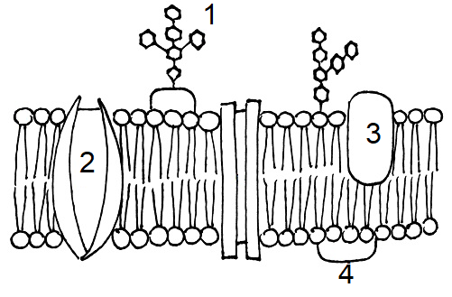
Protein channels cross the membrane to allow hydrophilic substances to pass through the membrane.
Which means of transport across a plasma membrane requires the molecule shown in the picture?

Identify the stage of mitosis of cells 1 and 2
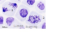
In cell 2, the chromatids are aligned on the equator (seen from above)- Metaphase. In cell 1, the chromatids are moving towards the poles - Anaphase.
Which of the following contributed to the acceptance of the fluid mosaic model of membrane structure of Singer and Nicholson in place of the original Davison-Danielli model?
I Hydrophobic membrane proteins
II Irregular sizes of membrane proteins
III Increased magnification of light microscopes.
IV Fluorescent antibody tagging.
The irregular sizes and insolubility of hydrophobic membrane proteins indicated that they could not be a surface layer as proposed by Davison-Danielli. This was confirmed by fluoresecent antibodies showing that proteins were both within and on the membrane.
Which of the following is the best description of an organelle?
The "wrong" answers are correct statements but are distractors, not the best description.
Refresh this page to try a new set of 20 multiple choice questions. The questions will be different next time you visit. Great revision.
 Twitter
  Facebook
  LinkedIn