
Topic 1: Cell biology 
This page contains multiple choice questions in the style of Paper 1 of the Biology exams.
They test the breadth of your knowledge of the understandings and skills about cell biology.
To spend more time reviewing the topic before answering these questions, use the revision resources.
Cell biology revision resources
This page lists the understandings and skills expected for Topic 1 and links to the sub-topic pages which contain detailed revision notes, activities and past paper style questions. Great for revision.
Learn from any mistakes. Every question has an examiner's explanation that appears when you check your answers.
Which cells are produced when a diploid cell divides by mitosis?
Mitosis is division of the nucleus into two genetically identical daughter nuclei in eukaryote cells.
A diploid cell will produce two diploid daughter cells in one division of mitosis.
The microscope image shows cells in a tissue sample taken from a growth suspected of being cancerous.
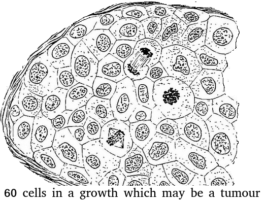
What is the best estimate of the mitotic index of this tissue?
Assume that there are exactly 60 cells (for simplicity).
Skill: Determination of a mitotic index from a micrograph
There are 3 cells in stages of mitosis.
So the mitotic index = 3 divided by 60 cells total.
Keeping it simple = 1 / 20 or 0.05
This box contains a lung waiting for a transplant operation.
![]()
What is special about the solution inside the box which surrounds the tissue?
Tissues and organs must be kept in a solution with the same osmolarity as the cytoplasm of the cells to prevent osmosis. If they were kept in pure water, osmosis would carry water into the cells and they would burst, causing damage to the cells. If the solution was hypertonic, the tissue would lose water (and gain ions).
The image below shows three structures as seen in an electron microscope.
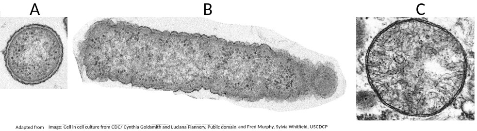
Which of the structures are prokaryote cells?
Students are expected to be able to recognise and draw the simple structure of Prokaryote cells.
There is no compartmentation in prokaryote cells, and as membranes can be seen in structure B (a mitochondrion) it is not a prokaryote.
Why is it that prokaryotes can divide by the simple process of binary fission, but eukaryotes have to divide by the more complex process of mitosis?
To explain how the structure of prokaryotes allows them to divide by binary fission you could mention:
- Prokaryotes have a single chromosome, eukaryotes have multiple chromosomes
- Prokaryotes have no nuclear membrane, which eukaryotes have.
How does compartmentalisation by their internal membranes benefit eukaryotic cells?
Eukaryote cells (approx. 100µm in diameter) are much larger than prokaryote cells (approx 1µm) and so the concentration of reactants in the cytoplasm would be more dilute if all the metabolism happened in the cytoplasm.
Specialist organelles, like mitochondria keep the enzymes for aerobic respiration in one place, which increases their concentration, and increases the rate of reactions.
The 'Cell theory' explains the nature of living things.
Which statement best describes Cell theory?
According to cell theory, living organisms are composed of cells.
Cells come from pre-existing cells and cells are the smallest using of life.
Human red blood cells are circular and 0.6 μm in diameter. A photograph of a red blood cell is shown as an illustration in a book with a diameter of 1.2mm. What is the magnification of the diagram?
Comment: Convert 1.2 mm into μm by multiplying x 1000 = 1200 μm (so that both units are the same). Then you can see that 0.6 x 2000 = 1200. Or use the formula Magnification = Image size/true size. If the photograph is larger than the cell, the magnification could not be 0.5x which would make it smaller. Eliminate obviously incorrect answers.
The image below is of Dracaena leaf upper epidermis cells.
Which of the following is the best estimate of the length (from top to bottom) of an epidermal cell?
Comment: The cells are approximately the same size as the scale bar.
This would make 70µm the closest estimate.
Why is a fungal hypha an exception to the cell theory?

A fungal hypha has many nuclei in a hypha but no cross walls to divide the hypha into cells.
What is the approximate length of the bacterial cell in the image?

The scale bar is 1 µm and the bacterium is approximately 3 times the length of the scale bar.
The diagram shows a typical eukaryotic plant cell. Which organelles are involved in supporting the cell and plant? I Cell wall II Cytoplasm III Nucleus IV Vacuole.

The cell and plant are supported by the turgor pressure of water in the vacuole acting on the rigid cell wall.
Which organelles are found in large numbers in secretory cells in animals? I Vesicles II Golgi Body III Mitochondria IV Rough endoplasmic reticulum.

Secretory cells synthesise proteins for exocytosis so have large numbers of mitochondria to supply energy, RER to synthesise the proteins for packaging into vesicles by the Golgi Body.
The image shows a transverse section of a plant cell seen using an electron microscope.
What is the main function of the large organelle (A) seen in the cell?

The organelle shown is the nucleus, it stores the genetic information, DNA and is the location of DNA replication and Transcription.
The image is a ribbon model of a channel protein. Where would this be found in a plasma membrane?

Channel proteins penetrate the membrane and have a central hydrophilic area (yellow in the diagram which is shown from above).
Which is the best description of the genetic code?
The genetic code is universal (the codons code for the same amino acid in all organisms) but there are a very few exceptions, mostly in Archaea. A mutation does not alter the genetic code, it alters the base sequence of DNA.
All eukaryote cells have differing combinations of the same types of organelles. How can this be explained?
Similarity in structure and function are taken as evidence of a common ancestor. All of the other statements are partially true but do not provide an explanation.
The theory of spontaneous generation has been disproved by Pasteur's experiment. Is there a point in evolution when spontaneous generation did occur?
The first cells must have arisen spontaneously from non-living matter, probably in volcanic vents, but the universal nature of cell ultrastructure and of the genetic code makes it likely that this only happened once.
Which of the following contributed to the acceptance of the fluid mosaic model of membrane structure of Singer and Nicholson in place of the original Davison-Danielli model?
I Hydrophobic membrane proteins
II Irregular sizes of membrane proteins
III Increased magnification of light microscopes.
IV Fluorescent antibody tagging.
The irregular sizes and insolubility of hydrophobic membrane proteins indicated that they could not be a surface layer as proposed by Davison-Danielli. This was confirmed by fluoresecent antibodies showing that proteins were both within and on the membrane.
How do integral proteins remain within the phospholipid bilayer?
Integral proteins have a hydrophobic centre that associates with the fatty acid inner layer of the bilayer and a hydrophilic head that associates with the phospholipid heads.
Refresh this page to try a new set of 20 multiple choice questions. The questions will be different next time you visit. Great revision.
 Twitter
  Facebook
  LinkedIn