
Topic 1: Cell biology 
This page contains multiple choice questions in the style of Paper 1 of the Biology exams.
They test the breadth of your knowledge of the understandings and skills about cell biology.
To spend more time reviewing the topic before answering these questions, use the revision resources.
Cell biology revision resources
This page lists the understandings and skills expected for Topic 1 and links to the sub-topic pages which contain detailed revision notes, activities and past paper style questions. Great for revision.
Learn from any mistakes. Every question has an examiner's explanation that appears when you check your answers.
What is the best definition of endosymbiosis?
Endosymbiosis is where a cell engulfs another cell and it continues to live inside the cell.
The engulfed cell provides something for the host cell, and gets something in return. Both cells benefit.
Which cells are produced when a diploid cell divides by mitosis?
Mitosis is division of the nucleus into two genetically identical daughter nuclei in eukaryote cells.
A diploid cell will produce two diploid daughter cells in one division of mitosis.
During interphase of the cell cycle what happens to the DNA in the nucleus?
Under the microscope there is little change during interphase.
However interphase is a very active phase of the cell cycle with many processes occurring in the nucleus and cytoplasm. (It is subdivided into G1, S, G2 phases of the cell cycle)
DNA replication occurs during the S-phase.
The microscope image shows cells in a tissue sample taken from a growth suspected of being cancerous.
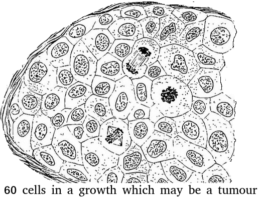
What is the best estimate of the mitotic index of this tissue?
Assume that there are exactly 60 cells (for simplicity).
Skill: Determination of a mitotic index from a micrograph
There are 3 cells in stages of mitosis.
So the mitotic index = 3 divided by 60 cells total.
Keeping it simple = 1 / 20 or 0.05
Identify the stage of mitosis for the two cells X and Y.
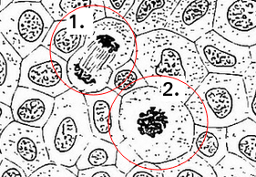
Skill: Identification of phases of mitosis in cells viewed with a microscope or in a micrograph
(prophase, metaphase, anaphase and telophase).
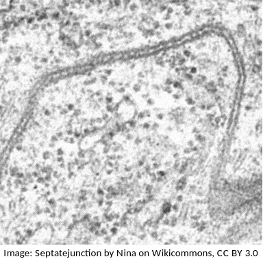 The photograph is from an electron microscope image of a membranes.
Parallel dark lines are clearly seen.
In electron micrography, heavy metal stains are often used to enhance contrast. DNA and Proteins often show as dark areas because these molecules can attach to the stain.
Which of the following interpretations is most similar to the proposal of the Daveson-Danielli model of membranes?
Students are expected to know how to use evidence from electron microscopy to support an idea about membranes. One illustration of this is the proposal of the Davson-Danielli model.
Further evidence lead to the falsification of the Davson-Danielli model and the proposal of the fluid mozaic model.
The image below shows three structures as seen in an electron microscope.
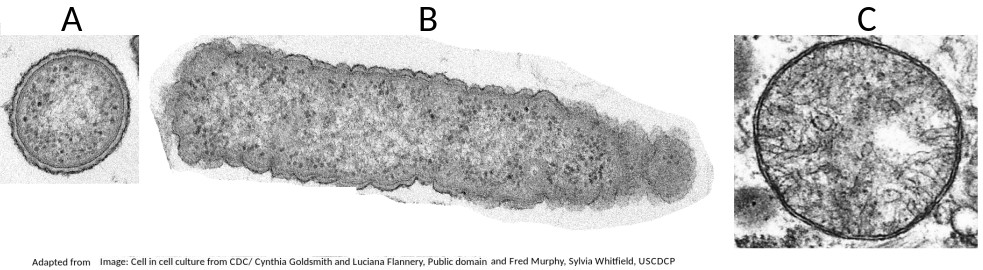
Which of the structures are prokaryote cells?
Students are expected to be able to recognise and draw the simple structure of Prokaryote cells.
There is no compartmentation in prokaryote cells, and as membranes can be seen in structure B (a mitochondrion) it is not a prokaryote.
The electron microscope image below shows an organelle found in eukaryote cells.
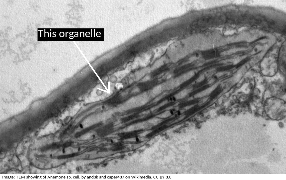
What is the name of the organelle?
Chloroplasts are distinctive because they have stacks of membranes inside, called grana, which hold the chlorophyll that absorbs light.
Why is it that prokaryotes can divide by the simple process of binary fission, but eukaryotes have to divide by the more complex process of mitosis?
To explain how the structure of prokaryotes allows them to divide by binary fission you could mention:
- Prokaryotes have a single chromosome, eukaryotes have multiple chromosomes
- Prokaryotes have no nuclear membrane, which eukaryotes have.
Cell theory covers most, but not all cases.
Which one of these statements is an exception?
Exceptions to cell theory are : multinucleated striated muscle the giant single celled Acetabularia algae?
Also, organisms consisting of only one cell carry out all functions of life in that cell. e.g. Paramecium, Chlorella.
Stargardts disease is vision loss caused by the death of both cone cells and rod cells in the part of the retina around the fovea. One potential treatment for Stargardts disease is the use of human embryonic stem cells.
What are the properties of these stem cells, which other cells don't have, that makes them so useful for this treatment?
Stem cells can divide and this help researchers to grow them in the lab.
They can differentiate along different pathways in embryonic development which makes stem cells useful for therapeutic uses (e.g. Stargart's disease) because they can be grown into many different tissues.
The blood cells below were imaged using an electron microscope.
The magnification is x3000 and the ruler measures the central cell as being 2 cm in diameter.
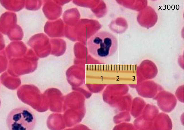
Estimate the actual size of this white blood cell.
Calculate specimen size using magnification?
First change the size measurement into µm units = 20000µm
Then divide by the magnification. 20000 / 3000 = 20 / 3 = 6.6 µm
The image below shows a eukaryotic cell.
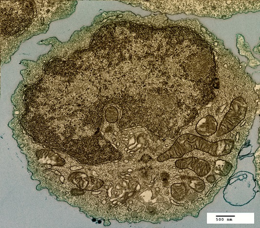
Which structure, visible in the image, could be used as evidence supporting endosymbiosis?
The mitochondria provide evidence supporting endosymbiosis because they have:
- a double membrane
- 70S ribosomes
- DNA
The image below is of Dracaena leaf upper epidermis cells.
Which of the following is the best estimate of the length (from top to bottom) of an epidermal cell?
Comment: The cells are approximately the same size as the scale bar.
This would make 70µm the closest estimate.
Which organelle in a eukarytotic animal cell synthesises proteins for exocytosis?

The RER synthesises proteins for exocytosis.
The diagram is of a plasma membrane. Which label corresponds to an extrinsic glycoprotein?

Extrinsic proteins are on the outside of the membrane, glycoproteins have carbohydrate prosthetic (side) groups (shown by the hexagonal shape).
The diagram is of a plasma membrane. Which label corresponds to a protein channel?
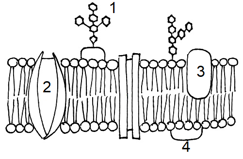
Protein channels cross the membrane to allow hydrophilic substances to pass through the membrane.
Which process is involved in white blood cells engulfing bacteria (arrowed in the photomicrograph)?

White blood cells engulf bacteria by endocytosis, the intake of solid particles by a cell membrane.
Louis Pasteur used sterile broth and swan necked flasks to disprove which theory?

Pasteur demonstrated that broth would go cloudy only when air was allowed to contact the broth, bringing microbes. He disproved Spontaneous Generation.
Which component of the plasma membrane varies in function between differentiated cells?
The protein component of the membrane varies in structure and function.
Refresh this page to try a new set of 20 multiple choice questions. The questions will be different next time you visit. Great revision.
 Twitter
  Facebook
  LinkedIn