
- Summary list for topic 1.2 Ultrastructure of cells
- Mindmaps
- Video tutorials
- Model answer
- Model answer
- Multiple choice questions
- 1.2 Ultrastrucure of cells quiz 1/1
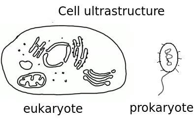 Eukaryote cells are larger than prokaryote cells and they have a more compartmentalised structure since endosymbiosis led to the creation of organelles. There are two basic types to draw, eukaryote and prokaryote cells. In this topic it's important to be able to recognise organelles in cells and to suggest cell functions depending upon cell structure.
Learn and test your biological vocabulary for 1.2 Ultrastructure of cells using these flash cards.


Summary list for topic 1.2 Ultrastructure of cells
- Know and be able to draw the simple structure of Prokaryote cells
- Know and draw the compartmentalised structure of Eukaryotic cells.
- Understand that the resolving power of electron microscopes is between 10µm and 1nm
whereas light microscopes resolve details between 1mm and 1µm.
Skills and applications
- Ability to identify organelles from microscope images of cells.
- Ability to explain how the composition of organelles will be different in cells with different functions,
(e.g. goblet cells which make mucus and palisade mesophyll cells which do photosynthesis). - Explain how the structure of prokaryotes allows them to divide by binary fission.
Mindmaps
This diagram summaries the main sections of topic 1.2 Cell ultrastructure.
Test if you can draw your own list or concept map from memory.


Video tutorials
How to draw a eukaryote animal cell diagram quickly in an exam.
How to draw a eukaryote animal cell in an exam. From David Faure on Vimeo.
How to identify organelles in electron microscope images and to explain how the structures relate to the cell's function.
Interpreting electron microscope images from David Faure on Vimeo.
This video help students to identify organelles and to make the link between cell structure and function.

Here is an example of a poor drawing of a prokaryotic cell.

This is what the teacher said about this diagram:
- The label lines are inaccurate.
- The flagellum is not in proportion to the cell.
- The nucleoid is incorrectly drawn.
- Cell wall and "cell membrane" are unclear.
- Missing/not labelled - plasmid, plasma membrane (not cell membrane), 70s ribosomes.
- Examiner hint - use clear solid lines and try to show size and shape in proportion.
Draw a better labelled diagram showing the correct structure of a prokaryote cell as it might be seen in an electron microscope image. (4)
.
.

Multiple choice questions
This self marking multiple choice quiz contains questions covering the topic.
START QUIZ!
The image below shows three structures as seen in an electron microscope.
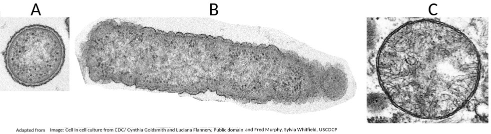
Which of the structures are prokaryote cells?
Students are expected to be able to recognise and draw the simple structure of Prokaryote cells.
There is no compartmentation in prokaryote cells, and as membranes can be seen in structure B (a mitochondrion) it is not a prokaryote.
The electron microscope image below shows three organelles found in an animal cell.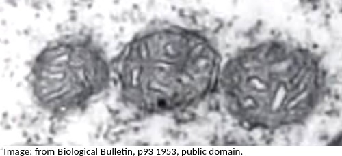
What is the name of the organelles?
Know how to identify the organelles in eukaryotes and draw their compartmentalised structure.
A mitochondrion (pleural = mitochondria) has an outer membrane and inner membrane folded into long thing 'flaps' called cristae.
The electron microscope image below shows an organelle found in both animal and plant cells.
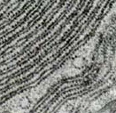
What is the name of the organelle?
Know how to identify the organelles in eukaryotes and draw their compartmentalised structure.
The rER has parallel membranes covered in dots, which are ribosomes, used for making proteins, for secretion from the cell.
The electron microscope image below shows a ciliated epithelial cell from the lungs.
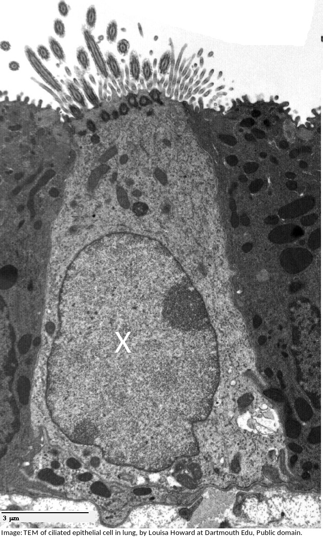
What is the name of the organelle labelled X?
The electron microscope image below shows an organelle found in eukaryote cells.
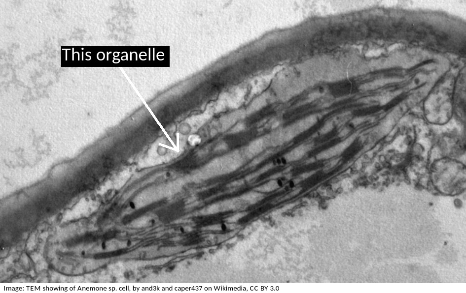
What is the name of the organelle?
Chloroplasts are distinctive because they have stacks of membranes inside, called grana, which hold the chlorophyll that absorbs light.
What are the structures labelled X and Y likely to be in this electron microscope image?
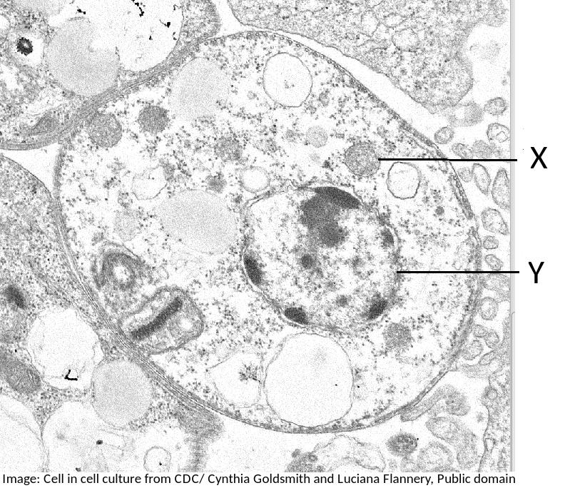
Students are expected to be able to identify organelles from microscope images of cells. The nucleus is distinctive because it is about 10µm in size, and it has black dots in it, chromatin, and sometimes one or more dark patches within the nuclear membrane. It also has a double membrane, not often easily visible.
If you found a eukaryote cell in an electron microscope image, and it contained a lot of rER, Golgi apparatus and many darkly stained vesicles, what do you think the function of the cell is most likely to be?
(eg. goblet cells which make mucus (a protien) will contain lots of rER and vesicles of musus, and palisade mesophyll cells which do photosynthesis will contain lots of chloroplasts)
Why is it that prokaryotes can divide by the simple process of binary fission, but eukaryotes have to divide by the more complex process of mitosis?
To explain how the structure of prokaryotes allows them to divide by binary fission you could mention:
- Prokaryotes have a single chromosome, eukaryotes have multiple chromosomes
- Prokaryotes have no nuclear membrane, which eukaryotes have.
This electron microscope shows a group of prokaryotes.
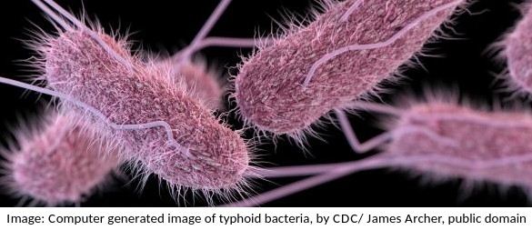
What structures are most likely to be found inside these cells?
Skill: you should know how to draw prokaryotic cells (with a cell wall, plasma membrane, cytoplasm, pili, flagella, 70s ribosomes and nucleoid.) and eukaryotic cells (free 80s ribosomes, rough endoplasmic reticulum (rER), lysosome, Golgi apparatus, mitochondrion and nucleus)
The electron microscope image below shows a cell.
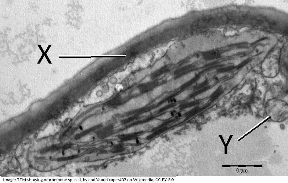
What are the organelles shown by the labels X & Y?
If you look closely at X, it points to the cell wall, outside the plasma membrane, it is close to the plasma membrane, but not touching the chloroplast.
The pale area below Y is the vacuole.
Organelle Y is a mitochondrion, you can tell this by its size, and the presence of membranes inside.
How does compartmentalisation by their internal membranes benefit eukaryotic cells?
Eukaryote cells (approx. 100µm in diameter) are much larger than prokaryote cells (approx 1µm) and so the concentration of reactants in the cytoplasm would be more dilute if all the metabolism happened in the cytoplasm.
Specialist organelles, like mitochondria keep the enzymes for aerobic respiration in one place, which increases their concentration, and increases the rate of reactions.
What is the term used to describe the smallest distance which two objects can be seen as separate objects in a microscope?
The resolving power, or resolution, is the ability to separate objects, to produce separate images of two objects.
Drag and drop activities
Test your ability to construct biological explanations using the drag and drop questions below.
Test your construction of biological knowledge using the drag and drop questions below.
Contrasting Eukaryote and Prokaryote cell structure.
Drag and drop the correct term into the gap to outline the differences between the cell structure of Prokaryotes and Eukaryotes.
plasmids 80s animal are always may nucleus 70s single multicellular mitochondria cell membrane chloroplasts naked protein membrane
Prokaryotic cells have a DNA whereas eukaryotic cells have chromosomes consisting of DNA and in a true membrane bound .
Prokaryotes are always celled whereas eukaryotes be .
Both eukaryotic cells and prokaryotes have ribosomes, but they are in prokaryotes and (larger) in eukaryotes.
Only eukaryotic cells have bound organelles such as and, in plant cells, .
Prokaryotes have cell walls; eukaryotic cells do not have a cell wall.
Explanation/Examiner hint. Contrast means to give the differences, using direct comparison.
Just for fun
Try this matching flashcards game.
If you can't see the content below, please click this link Ultrastructure of cells Quizlet matching game.
Alternatively try this Arcade game to practise the basics. Arcade game "Ultrastructure of cells".
 Twitter
  Facebook
  LinkedIn