
 This simple revision quiz contains questions to help you revise the topic A2.2 Cell Structure
This topic is all about the use of microscopes, preparation of slides and the structure of prokaryote and eukaryote cells.
The electron microscope image below shows a scale bar marked with 100µm.
The large 'goblet cell' in the centre is producing mucous which will protect the surface of the epithelium.
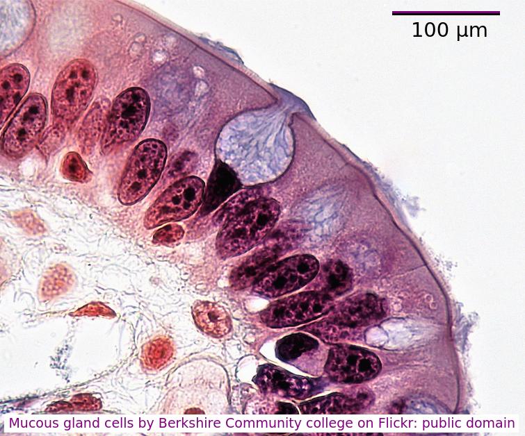
What is the diameter of the goblet cell?
Accurately, measure the scale bar length in mm, measure the diameter of the cell, in mm
divide cell diameter by scale bar and multiply by 100µm.
You can often estimate the size using the scale bar and your thumb or a pen.
Human red blood cells are circular and 0.6 μm in diameter. A photograph of a red blood cell is shown as an illustration in a book with a diameter of 1.2mm. What is the magnification of the diagram?
Comment: Convert 1.2 mm into μm by multiplying x 1000 = 1200 μm (so that both units are the same). Then you can see that 0.6 x 2000 = 1200. Or use the formula Magnification = Image size/true size. If the photograph is larger than the cell, the magnification could not be 0.5x which would make it smaller. Eliminate obviously incorrect answers.
The diameter of this field of view under a microscope at X400 magnification is 250 μm.
The image below shows Dracaena leaf upper epidermis cells.
Which is the best estimate of the width (from left to right) of an epidermal cell?
Comment: There are approximately 8 cells across a diameter so 250/8 = 33 μm.
Beware of the units, mm are 1000 times bigger than µm.
The image below is of Dracaena leaf upper epidermis cells.
Which of the following is the best estimate of the length (from top to bottom) of an epidermal cell?
Comment: The cells are approximately the same size as the scale bar.
This would make 70µm the closest estimate.
What is the structure of the genetic material found in a mitochondrion?
Mitochondrial DNA is a single helical molecule, not associated with protein and circular in shape. The same as prokaryote nucleoid DNA.
The image below shows a eukaryotic cell.
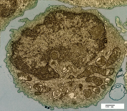
Which structure, visible in the image, could be used as evidence supporting endosymbiosis?
The mitochondria provide evidence supporting endosymbiosis because they have:
- a double membrane
- 70S ribosomes
- DNA
Which organelles in a plant cell are believed to have originated as free-living prokaryotic cells?

Both the mitochondria and the chloroplast in plant cells are thought to have been free-living prokaryotes which evolved in a symbiotic relationship with a eukaryotic cell.
What is the best definition of endosymbiosis?
Endosymbiosis is where a cell engulfs another cell and it continues to live inside the cell.
The engulfed cell provides something for the host cell, and gets something in return. Both cells benefit..
Which of the following can be regarded as advantages of the multicellular condition?
In the multicellular condition, the organism can generally be larger and cells can specialise to form tissues and organs. Multicellular organisms have the same organelles as single celled and cephalisation is the concentration of the sensory system at the anterior end of some Animalia.
Complete the sentences by filling in the gaps.
differentiation unicellular mutation multicellular environment expression specialised
Cell allows the development of tissues in organisms. It is caused by changes in gene usually in response to the internal or external .
Cells differentiate in response to changes in gene expression, usually triggered by the environment.
Which of the following could be used to distinguish between a prokaryote and a eukaryote cell?
All prokaryotic cells have a cell wall, plant eukaryotic cells have a cell wall. If there is no cell wall, the cell must be an eukaryotic animal cell. Prokaryotes have smaller 70s ribosomes than eukaryotes (80s). Both have DNA and both may have flagellae.
This question compares and contrasts prokaryote and eukaryote ultrastructure. Drag and drop the correct word or words into the space provided.
ribosomes Eukaryotic animal cells mitochondria flagella 80s cell walls
Both eukaryotic cells and prokaryotes have , but they are 70s in prokaryotes and (larger) in eukaryotes. Both cell types may have .
Only eukaryotic cells have .
Both plant eukaryotic cells and prokaryotes have . do not have a cell wall.
Compare and contrast - similarities and differences in one sentence.
Other points can include:
Prokaryotes are single celled whereas eukaryotes may be multicellular.
The image shows a transverse section of a plant cell seen using an electron microscope.
What is the main function of the large organelle (A) seen in the cell?

The organelle shown is the nucleus, it stores the genetic information, DNA and is the location of DNA replication and Transcription.
Which organelles are found in large numbers in secretory cells in animals? I Vesicles II Golgi Body III Mitochondria IV Rough endoplasmic reticulum.

Secretory cells synthesise proteins for exocytosis so have large numbers of mitochondria to supply energy, RER to synthesise the proteins for packaging into vesicles by the Golgi Body.
Which organelle is visible in an electron microscope but not in a light microscope?

The ribosome is too small to be seen in the electron microscope, the other organelles were seen in the light microscope before the electron microscope was invented.
The image is of a prokaryotic cell. Which feature defines the cell as prokaryotic?
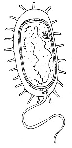
Naked DNA in a single molecule shaped like a loop (nucleoid) defines a cell as prokaryotic.
The diagram shows a typical eukaryotic plant cell. Which organelles are involved in supporting the cell and plant? I Cell wall II Cytoplasm III Nucleus IV Vacuole.

The cell and plant are supported by the turgor pressure of water in the vacuole acting on the rigid cell wall.
The electron microscope image below shows a cell.
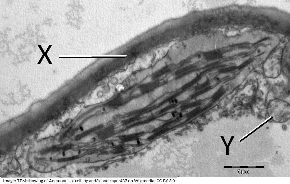
What are the organelles shown by the labels X & Y?
If you look closely at X, it points to the cell wall, outside the plasma membrane, it is close to the plasma membrane, but not touching the chloroplast.
The pale area below Y is the vacuole.
Organelle Y is a mitochondrion, you can tell this by its size, and the presence of membranes inside.
Between X and Y is the chloroplast which is larger than the mitochondrion and has a different internal membrane structure.
This electron microscope image shows a group of prokaryotes.
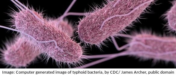
What structures are most likely to be found inside these cells?
Prokaryotes have a cell wall (of peptidglycan) and all cells have a membrane. They do not have membrane bound organelles.
What are the structures labelled X and Y likely to be in this electron microscope image?
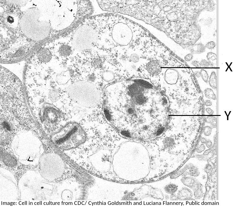
The nucleus is distinctive because it is large, and it has black dots in it of chromatin, and sometimes one or more dark patches within the nuclear membrane (nucleoli). It also has a double membrane, not often easily visible.
The mitochondrion is smaller and has convoluted inner membranes.
 Twitter
  Facebook
  LinkedIn